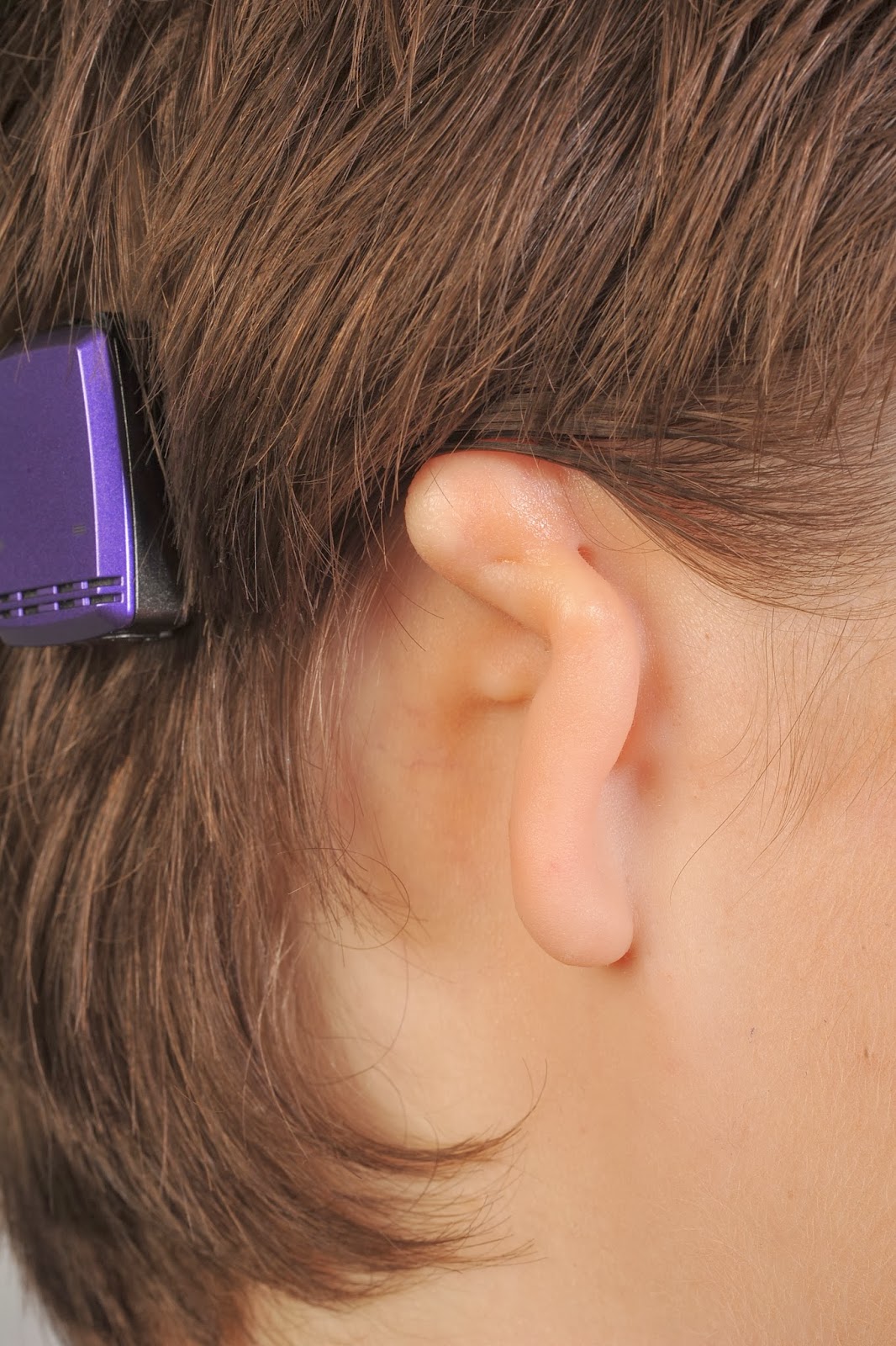
Explanations follow the photos:
These first two photos are what the ear looked like right after it was sewn up. You can see more definition here than in my current swelled state. The finished product will be much more defined than these photos show. I saw a picture of a little boy who had surgery 6 months ago. It looked like he had had a canal built, but I was amazing to hear that he hadn't, and that my ear will look similar when it's all healed (it will probably take my ear 1 to 1 and a half years to completely heal). Also, if you notice the three little stitches near the top of the ear, those were put in to close tiny holes in the skin (that skin was from my microtia ear) from where the microtia ear had tiny little holes/dips in it. It's hard to see from this angle, but you can see the top of the 3 holes in the top front area of the ear in the following photo:
Here's the carved cartilage! The black lines are all the stitches holding the pieces together. You can see in the picture on the right that Dr. Griffiths stacks layers of cartilage up to gain the projection for the ear so that he can accomplish the elevation of the ear in the same surgery as the initial placement of the cartilage ear framework. My existing ear is interesting in features. My outer rim gets pretty flat after the top portion, which is reflected in this new ear. Apropos, the highest point on my existing ear is not the rim on the side of the ear, but the spot inside the rim just before the ear goes down towards the canal...thus the large piece of cartilage Dr. Griffiths added to that area on this ear. There are a lot of other 3D nuances in the carving, but it is a little difficult to see in the photos due to the angles and the fact that the cartilage varies in color naturally, so it's hard to know when it's the cartilage or the depth causing a change in color.
More questions? Feel free to leave a comment and I can see if I know the answer!
~Katie


No comments:
Post a Comment